
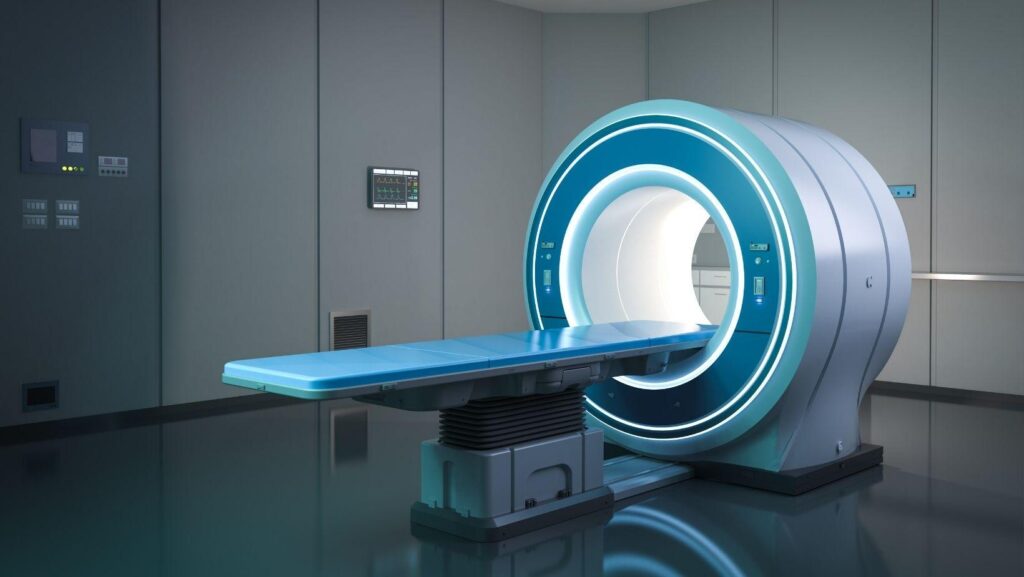
Dental implants require more than a visual examination. CT scans and 3D imaging show your jawbone, teeth, nerves, and sinus structures in detail, giving your dental team information that standard X-rays cannot provide. This kind of digital planning is a routine part of preparing for dental implants in Wilmington, helping ensure the treatment plan fits your specific anatomy.
By using 3D imaging, you can receive a treatment plan based on your actual bone volume, anatomy, and implant needs. Your dentist can evaluate the surgical site, identify potential challenges, and plan the implant's position, size, and angle before treatment begins.
Digital planning supports more precise placement. It helps your team assess practical factors such as bone availability, surgical access, and possible need for additional procedures.
Diagnostic Imaging Fundamentals
Your dentist uses imaging to measure available bone, identify vital structures, and select an implant position before surgery. The choice between conventional X-rays and CBCT depends on the procedure’s complexity, the anatomy involved, and the amount of detail required.
Cone Beam CT Versus Conventional Dental X-Rays
Conventional dental X-rays produce two-dimensional images. Periapical and panoramic views can show teeth, roots, general bone levels, and some jaw conditions, but they compress three-dimensional anatomy into a flat image.
This can obscure the cheek-to-tongue width of the ridge, overlap nearby structures, and make distances difficult to judge accurately. CBCT creates a three-dimensional scan using a cone-shaped X-ray beam.
Your dentist can examine cross-sectional, panoramic, and axial views to assess bone height, width, and contour. The location of the mandibular nerve and mental foramen, sinus position and possible sinus abnormalities, adjacent roots, impacted teeth, lesions, and available space for the implant and restoration are also visible.
CBCT exposes you to radiation, although the dose varies by scanner and field of view. Your clinician should prescribe it when the additional information can affect diagnosis, implant positioning, or surgical safety.
Creating Three-Dimensional Anatomical Views
CBCT software reconstructs your jaw from multiple images and allows your clinician to review the anatomy in thin slices. These views show the ridge from different directions, helping reveal concavities, narrow areas, and changes in bone volume that a panoramic image may not display.
Your dentist can use digital measurements to compare the proposed implant’s length and diameter with the available bone. The software also helps identify a safe distance from nerves, adjacent roots, and the maxillary sinus.
When combined with digital impressions or an intraoral scan, the CBCT dataset can support prosthetically guided planning. This positions the implant according to the planned crown.
The scan does not measure bone strength directly or remove the need for a clinical examination. Image quality can also be affected by patient movement and metal artifacts, so your clinician must interpret the scan alongside your medical history, examination findings, and restorative plan.
Anatomical Assessment Before Surgery
A CT scan or CBCT scan gives your dentist a three-dimensional view of the proposed implant site. This helps identify whether you have enough supporting bone and shows nearby structures that could affect implant position, size, or surgical approach.
Measuring Bone Volume And Density
Your scan allows the dental team to measure bone height, width, and shape at the planned implant site. These measurements help determine whether the implant can fit securely or whether you may need bone grafting before placement.
The images also show areas of bone loss that two-dimensional X-rays may not reveal clearly. Your dentist can assess the ridge from multiple angles, identify thin sections, and choose an implant diameter and length that match your anatomy.
CBCT does not measure bone density in exactly the same way as a medical CT scan. Your dentist combines the scan with clinical findings.
Bone quality affects initial stability and may influence implant placement, healing time, and loading plans.
Locating Nerves, Sinuses, And Adjacent Roots
Three-dimensional imaging helps your dentist locate the inferior alveolar nerve, mental nerve, and other important structures near the implant site. In the upper jaw, the scan shows the maxillary sinus and can reveal limited space, sinus expansion, or conditions that may require additional evaluation.
The scan also displays neighboring tooth roots, impacted teeth, and other anatomical features. Your dentist can use this information to maintain safe distances and select an implant angle that avoids contact with these structures.
If the scan identifies a potential risk, your treatment plan may change. Options can include a shorter or narrower implant, a different placement position, staged grafting, or referral to an appropriate specialist.
Digital Implant Placement Planning
CT and 3D imaging let you evaluate bone volume, identify nearby nerves and sinuses, and plan implant placement against the intended crown or bridge. This information helps you select an appropriate implant position and create a guide that transfers the digital plan to surgery.
Selecting Implant Size And Position
Your clinician reviews the CBCT scan to measure available bone height, width, and density. The plan must maintain safe distances from the inferior alveolar nerve, sinus cavity, adjacent tooth roots, and other anatomical structures.
If the scan shows insufficient bone, you may need grafting or an alternative treatment plan before implant placement. The implant position should support the final restoration, not simply occupy the largest area of available bone.
Digital planning software allows your clinician to place a virtual implant and evaluate its diameter, length, angulation, and depth in relation to the planned crown. This prosthetically driven approach helps position the implant where it can support chewing forces and allow suitable emergence through the gum.
Your clinician may also combine the CBCT scan with a digital impression or intraoral scan. This merges anatomical information with the planned tooth shape, improving coordination between implant placement and the restoration.
Designing Surgical Guides
After selecting the implant position, your clinician can use planning software to design a patient-specific surgical guide. The guide fits over your teeth, gums, or supporting bone and contains sleeves that control the drill path.
It transfers the planned entry point, angle, and depth to the surgical site. A tooth-supported guide often provides stable positioning when enough natural teeth remain.
A tissue-supported or bone-supported guide may suit patients with extensive tooth loss, although the clinician must account for movement and support during surgery. The design also considers access, guide thickness, fixation pins, and the sequence of drilling instruments.
Before fabrication, your clinician checks the digital plan for conflicts with the planned restoration and surrounding anatomy. Guided surgery can improve placement control, but it does not replace clinical judgment.
The clinician still verifies fit, visibility, and anatomy during the procedure.
Clinical Benefits And Practical Considerations
Three-dimensional imaging helps you evaluate bone anatomy, nearby structures, and restorative requirements before implant surgery. Selecting the appropriate scan also matters because you should balance diagnostic value with radiation exposure, cost, and the clinical question.
Improving Accuracy And Predictability
A CBCT scan provides volumetric images that show bone height, width, and angulation more clearly than conventional two-dimensional radiographs. You can assess the position of the mandibular canal, mental foramen, maxillary sinus, and adjacent roots before selecting an implant site.
This information supports safer planning and helps you identify whether bone grafting or sinus augmentation may be necessary. You can combine the scan with digital impressions or intraoral scans to plan the implant in relation to the proposed crown.
This restorative perspective helps you select implant position, depth, and angulation based on both available bone and functional demands. Virtual planning may also support a surgical guide, although the guide does not eliminate the need to verify anatomy during surgery.
Radiation Exposure And Imaging Selection
You should not obtain CBCT imaging automatically for every dental implant case. Start with the clinical examination and appropriate two-dimensional radiographs.
Use CBCT when cross-sectional or three-dimensional information could change diagnosis, treatment, or surgical risk. For example, complex anatomy, limited bone volume, proximity to vital structures, or previous grafting may justify a scan.
CBCT generally delivers less radiation than medical CT, but exposure varies with the machine, field of view, resolution, and scan settings. Use the smallest field of view and lowest suitable dose that answer the clinical question.
A larger field or higher resolution may provide more detail but also increases exposure. Your clinician should explain why imaging is needed and consider prior scans to avoid unnecessary repeat examinations.