
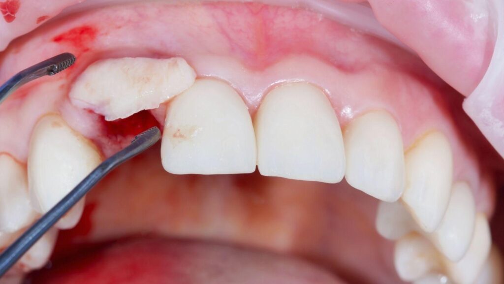
When a damaged or failing tooth needs removal, your dentist may recommend placing bone-grafting material in the extraction socket during the same visit. This combined approach can help preserve the shape and volume of the jaw while the site heals.
You may receive an extraction and bone graft in one appointment, but your dentist must confirm that the treatment suits your bone condition, infection status, and future tooth-replacement plan.
When planned well, this step can make bone grafting for dental implants in Greenville, SC more straightforward down the line by preserving bone volume from the start. You'll learn how the procedure works, what recovery involves, and how the graft can support a future dental implant or other restoration.
Why Extraction Sites Need Bone Support
Removing a tooth eliminates the stimulation that helps maintain the surrounding jawbone. Without preservation, the socket commonly loses width and height during healing, which can affect gum contours, neighboring teeth, and future implant placement.
Post-Extraction Bone Changes
After extraction, your body fills the socket with a blood clot, followed by healing tissue and new bone. The bone that once surrounded the tooth root then undergoes normal remodeling because it no longer receives pressure from chewing.
The outer wall, especially the thin facial or cheek-side plate, often resorbs first. This remodeling can reduce the ridge’s width and vertical height.
The amount varies with the tooth’s location, socket-wall thickness, infection, trauma during extraction, smoking, and individual healing factors. A smaller ridge may leave less bone for an implant and can change the soft-tissue shape around a future crown or bridge.
Bone loss does not necessarily cause symptoms, so you may not notice the change while the gum appears healed. Your dentist evaluates the socket clinically and, when needed, with dental imaging to determine whether preservation or later bone reconstruction would support your treatment plan.
Goals of Ridge Preservation
Ridge preservation places graft material into the extraction socket after the tooth is removed. The material acts as a scaffold or space maintainer while your body forms new bone and gradually remodels the graft.
Depending on the case, your dentist may also use a resorbable membrane, collagen plug, or sutures to protect the site during early healing. The primary goal is to limit collapse of the socket’s shape, particularly along the facial wall.
Preservation does not guarantee that no bone will change, and it does not replace careful extraction technique or later grafting when substantial bone is missing. Your dentist selects the graft material and timing based on the socket anatomy, infection status, remaining bone, and planned tooth replacement.
How the Combined Treatment Is Performed
Your dentist first confirms whether immediate grafting is appropriate, then removes the tooth and cleans the socket before placing graft material. The procedure may preserve the socket for future implant treatment, but the exact steps depend on infection, bone quality, gum health, and the condition of the surrounding tissues.
Clinical Examination and Imaging
Your dentist examines the tooth, gums, bite, and nearby teeth. They check for swelling, drainage, gum disease, looseness, and signs of infection that could affect healing.
You also review your medical history, medications, smoking or vaping habits, and any conditions that may influence bone repair. Dental X-rays show the tooth roots and surrounding bone.
For complex cases, cone-beam computed tomography (CBCT) creates a three-dimensional view of the jaw, nasal cavity, and sinus. This helps your dentist assess socket-wall damage, bone thickness, and the position of nerves or other structures.
Your dentist uses these findings to decide whether to graft immediately, delay treatment, or combine grafting with implant placement. An active, uncontrolled infection, major bone loss, or inadequate stability may require staged treatment.
Tooth Removal and Site Preparation
Your dentist numbs the area with local anesthetic. If necessary, sedation can help you remain comfortable.
The dentist removes the tooth as carefully as possible, often using instruments that loosen the tooth gradually rather than applying excessive force. This approach helps protect the thin bone walls and gum tissue around the socket.
After removal, the dentist inspects the socket for root fragments, infection, and defects. They remove inflamed tissue and rinse the area with sterile solution.
The socket must contain healthy, bleeding bone before graft material is placed. If the socket has lost a wall or contains substantial infection, your dentist may postpone grafting or use additional regenerative techniques.
Immediate implant placement remains a separate decision and requires enough healthy bone to hold the implant securely.
Graft Placement and Protective Materials
Your dentist fills the cleaned socket with graft material selected for your defect and treatment plan. Materials may come from a donor source, an animal-derived source, a synthetic substitute, or, less commonly, your own bone.
The graft supports the space while your body gradually forms new bone around the particles. The dentist places enough material to support the socket without overpacking it.
A resorbable collagen membrane may cover the graft, especially when the socket wall is damaged or the graft needs protection from soft-tissue pressure. The dentist then closes the gum with sutures or uses a protective dressing.
You receive instructions to avoid disturbing the site, including limiting pressure, vigorous rinsing, and smoking. Follow-up visits allow your dentist to check healing and determine when the area has enough bone for a future implant.
Healing, Recovery, and Follow-Up
Your gum tissue usually begins closing over the extraction and graft site within days, while the graft gradually integrates with your jawbone over several months. Careful cleaning, activity limits, prescribed medications, and scheduled examinations help protect the graft during this process.
Expected Recovery Timeline
During the first 24 hours, mild bleeding, swelling, tenderness, and limited jaw movement are common. Swelling often increases during the first 48–72 hours before it gradually improves.
You may notice soreness for several days, especially when chewing or opening your mouth. During the first one to two weeks, the gum tissue continues to heal.
Follow your surgeon’s instructions about removing or protecting any dressing, sutures, or temporary tooth replacement. Your clinician may check the site within one to two weeks.
The graft itself generally requires three to six months to mature sufficiently for implant placement, although the timing varies. Your surgeon may use follow-up examinations and CBCT imaging to confirm that the bone has healed adequately.
Smoking, certain medical conditions, infection, and extensive grafting can lengthen this timeline.
Aftercare Instructions
Signs That Require Prompt Attention
Contact your dental surgeon promptly if bleeding remains heavy after sustained pressure, swelling continues to worsen after the first few days, or pain becomes severe despite the recommended medication. A fever, foul taste, persistent bad breath, pus, or increasing redness can indicate infection.
Call promptly if you notice the graft material repeatedly coming out, an opening in the wound, exposed bone, numbness that does not improve, or difficulty swallowing. Seek emergency care for trouble breathing, rapidly spreading facial or neck swelling, uncontrolled bleeding, or signs of a serious medication reaction.
Do not wait for a scheduled appointment when symptoms change suddenly. Your surgeon can determine whether you need an examination, medication adjustment, additional cleaning, or other treatment.
Planning for Future Tooth Replacement
Your replacement plan affects whether grafting occurs during extraction, after healing, or with implant placement. A clinical examination and 3D imaging help your dentist assess infection, bone volume, gum tissue, implant position, and the timing of your final restoration.
Dental Implant Timing
Your dentist may place an implant during extraction when the socket has suitable bone, the infection is controlled, and the implant can achieve firm initial stability. A small graft may fill a gap around the implant or support the outer socket wall.
Immediate placement does not suit every tooth, particularly when infection, bone loss, thin tissue, or limited implant stability is present. If your dentist removes the tooth and grafts the socket first, you may wait several months for healing before implant placement.
After placement, the implant commonly needs about 3–6 months to integrate with the surrounding bone before your dentist attaches the final crown. Significant grafting or complex healing can extend the full treatment time to approximately 9–15 months.
Ask whether the graft aims to preserve the socket, support an immediate implant, or rebuild bone for a later implant. These goals involve different materials, procedures, and healing schedules.
Factors Affecting Long-Term Results
Your long-term result depends on the amount and quality of bone, the condition of your gums, implant positioning, and control of infection. Smoking, poorly controlled diabetes, untreated gum disease, and inadequate oral hygiene can interfere with healing and increase implant complications.
Your dentist may recommend a CBCT scan to measure bone width and height and to locate nearby nerves or sinus spaces. The plan should also account for your bite, tooth position, gum thickness, and the appearance of the final crown.
A graft can preserve volume. However, it cannot guarantee that an implant will remain possible without further treatment.
Ask about the graft material, expected healing time, temporary tooth options, and the conditions required before implant placement. Follow your aftercare instructions carefully, avoid disturbing the graft, attend review appointments, and maintain regular professional cleanings.